
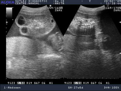
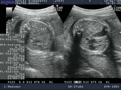
ACARDIAC PARABIOTIC TWIN
Twin pregnancy with 27 weeks of gestation shows an ill formed mass consisting of partially developed lower half of fetal body, lower limbs and lumber spine. Fetal head, upper limbs and upper part of trunk was not formed. Multiloculated dorsal cystic hygroma was also present.The second twin was normal at the time of ultrasound.
Twin pregnancy with 27 weeks of gestation shows an ill formed mass consisting of partially developed lower half of fetal body, lower limbs and lumber spine. Fetal head, upper limbs and upper part of trunk was not formed. Multiloculated dorsal cystic hygroma was also present.The second twin was normal at the time of ultrasound.
Double click to edit
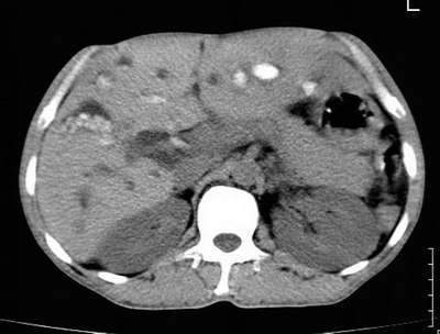
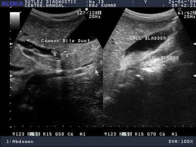
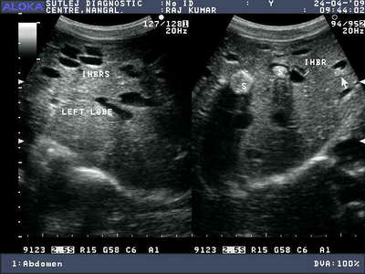
RECURRENT PYOGENIC CHOLANGITIS
Liver shows dilated 1st and 2nd order biliary redicals. Multiple echogenic lesions with and without acoustic shadowing are seen in CBD nad IHBR (s/o intraductal sludge and calculi).
Liver shows dilated 1st and 2nd order biliary redicals. Multiple echogenic lesions with and without acoustic shadowing are seen in CBD nad IHBR (s/o intraductal sludge and calculi).
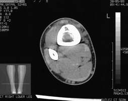
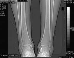
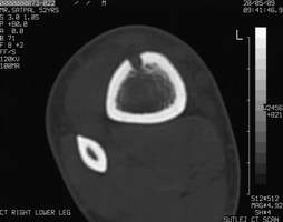
Adamantinoma of tibia is a rare tumor and till date only 200 cases have been reported in the world. The X-ray shows a well defined osteolytic lesion predominantly intracortical in the lower part of tibia. CT scan confirms these findings
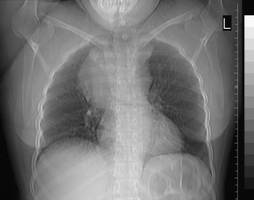
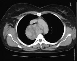
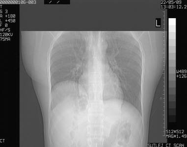
The CT topogram reveals a superior mediastinal mass with lobulated margins extending laterally mainly on right. CT sections show a lobulated heterogeneous mass in the mediastinum, posterior to the trachea and esophagus extending from the left paratracheal region superiorly crossing towards the right side posterior to the trachea and esophagus inferiorly. Superiorly it extended towards the left side of the posterior mediastinum and occupied the left paratracheal region, compressing and displacing the trachea
ADAMANTINOMA TIBIA
INTRATHORACIC GOITER
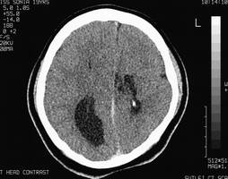
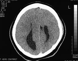
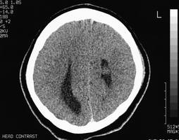
CT sections showning Agenesis of the corpus callosum with gray-matter heterotopia with right hemimegalencephaly
AGENESIS OF CORPUS CALLOSUM

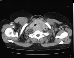
INTRATHORACIC KIDNEY
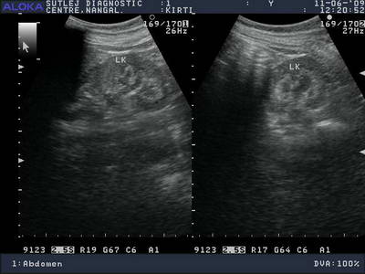
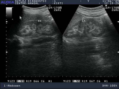
MEDULLARY NEPHROCALCINOSIS
CONGENITAL DIAPHRAMATIC
HERNIA
HERNIA
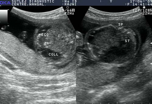
MECONIUM PERITONITIS
Meconium peritonitis in a fetus of 23 weeks gestational age. Multiple calcifications with a multiseptate fluid collections are seen in the abdomen
